
Eye Photography (retina / posterior segment of the eye)
The fundus camera is used to take photographic images of the posterior segments of the eye (retina).
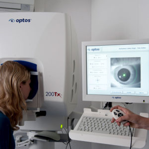
OPTOS PHOTOGRAPHY (ULTRA WIDE ANGLE)
The Optos device generates digital retinal images using a special laser scanning technology; pupil dilation is not necessary. With an Optos image, sections of the retina can be imaged up to 200° (82%) by using a mirror technology (conventional maximum 50°). Since many changes are first visible in the periphery, this examination is a simple and fast way to visualize and document the retina without exposing the eye to much light.
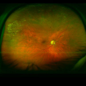
Vascular occlusions, retinal detachments, diabetic retinal changes, tumors and retinal holes can be detected.
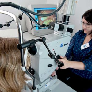
ZEISS CAMERA (20°/30° AND 50°)
The Zeiss camera is a device that takes photos of the retina with flashes like a reflex camera.
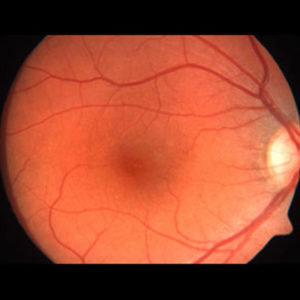
Due to the high resolution an exact documentation of the visual center (macula) and the optic nerve (papilla) is possible.
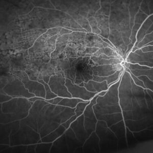
FLUORESCEIN AND INFRACYANINE GREEN ANGIOGRAPHY
The HRA Spectralis, Optos and Zeiss camera devices can be used to perform fluorescein and infracyanine green angiography. A contrast medium is injected intravenously. During the following 10 minutes, photographs of the retina are taken. This examination serves to precisely determine pathological changes in the retina, its vessels and neighboring structures. These findings are important in order to assess the treatment options and to be able to carry out a targeted therapy.