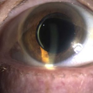
Anterior segment photography
SLIT LAMP / BIOMICROSCOPIC EXAMINATION
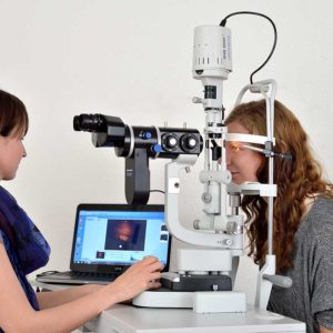
The slit lamp is a microscope with a swiveling light source. The lightning of the slit lamp is sharply defined and can be changed in shape and diameter. The magnification range of the microscope is variable and thus allows examination of all eye segments. With the lowest magnification (6-times) the eyelids and the corresponding posterior eye segments are examined. For the examination of the tear film, cornea, conjunctiva, anterior chamber of the eye, lens and vitreous body, magnifications of 10 to 40-times are set. The slit lamp is an important diagnostic instrument and is used by ophthalmologists almost without exception during the examination.
The patient sits opposite the doctor and positions the head on a chin rest. The ophthalmologist illuminates the eye and looks stereoscopically through the microscope. In order to better detect corneal defects, sometimes colored drops (fluorescein) are added to the conjunctival sac. This leads to a slight yellowish discoloration of the eye, which can be washed out.
To allow the doctor to assess the posterior segments of the eye, the pupils must be dilated with special eye drops (mydriatic). Afterwards, vision is impaired for about four hours, which is why one should NOT drive during this time.
With the slit-lamp camera, almost all observed changes and findings can be documented photographically.